
Description
A urinalysis is a group of manual and/or automated qualitative and semi-quantitative tests performed on a urine sample. A routine urinalysis usually includes the following tests: color, transparency, specific gravity, pH, protein, glucose, ketones, blood, bilirubin, nitrite, urobilinogen, and leukocyte esterase. Some laboratories include a microscopic examination of urinary sediment with all routine urinalysis tests. If not, it is customary to perform the microscopic exam, if transparency, glucose, protein, blood, nitrite, or leukocyte esterase is abnormal.
Purpose:
Routine urinalyses are performed for several reasons:
- general health screening to detect renal and metabolic diseases
- diagnosis of diseases or disorders of the kidneys or urinary tract
- monitoring of patients with diabetes
In addition, quantitative urinalysis tests may be performed to help diagnose many specific disorders, such as endocrine diseases, bladder cancer, osteoporosis, and porphyrias (a group of disorders caused by chemical imbalance). Quantitative analysis often requires the use of a timed urine sample. The urinary microalbumin test measures the rate of albumin excretion in the urine using laboratory tests. This test is used to monitor the kidney function of persons with diabetes mellitus. In diabetics, the excretion of greater than 200 ?g/mL albumin is predictive of impending kidney disease.
How the test is performed:
A urine sample is needed. Your health care provider will tell you what type of urine sample is needed.
24-hour urine collection
The urine 24-hour volume test measures the amount of urine produced in a day
24-hour urine sample is needed.
- On day 1, urinate into the toilet when you get up in the morning.
- Afterwards, collect all urine in a special container for the next 24 hours.
- On day 2, urinate into the container when you get up in the morning.
- Cap the container. Keep it in the refrigerator or a cool place during the collection period.
- Label the container with your name, the date, the time of completion, and return it as instructed.
For an infant, thoroughly wash the area around the urethra. Open a urine collection bag (a plastic bag with an adhesive paper on one end), and place it on the infant. For males, place the entire penis in the bag and attach the adhesive to the skin. For females, place the bag over the labia. Diaper as usual over the secured bag.
This procedure may take a couple of attempts — lively infants can move the bag, causing the urine to be absorbed by the diaper. Check the infant frequently and change the bag after the infant has urinated into it. Drain the urine from the bag into the container provided by your health care provider.
Deliver it to the laboratory or your health care provider as soon as possible upon completion.
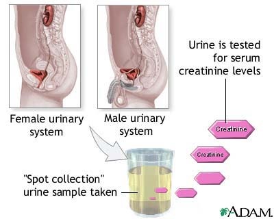
Clean catch urine specimen
A clean catch is a method of collecting a urine sample for various tests, including urinalysis and urine culture.
To obtain a clean-catch urine sample, boys and men should wipe the head of the penis clean. Girls and women need to wipe between the vagina “lips” (labia) with soapy water and rinse well. Your doctor may give you a special clean-catch kit that contains a cleansing solution and sterile wipes.
To perform this test, first urinate a small amount into the toilet bowl to clear the urethra of any contaminants. Then, collect a sample of urine in a clean or sterile container. About 1 – 2 ounces of urine is needed for a test. Remove the container from the urine stream without stopping the flow. You may finish urinating into the toilet bowl. Take the sample to the lab.
For infants, the genital area is cleaned and dried, and then a collection device is attached to collect the urine. If you are asked to collect the urine, be sure the collection device is attached securely to prevent leakage. After your baby has urinated, the urine (at least 20 cc) is placed in a sterile container.
Do not use antiseptics, as they may prevent bacteria from growing during the culture.
Normal Values:
- Volume: 600 to 2500 mL in 24 hours
- Color: Pale yellow to amber
- Appearance: clear to slightly hazy
- Specific gravity: 1.005 to 1.025 with a normal fluid intake
- pH: 4.5 to 8
- Glucose: negative
- Ketones: negative
- Blood: negative
- Protein: negative
- Bilirubin: negative
- Nitrate for bacteria: negative
- Casts: negative, occasional hyaline casts
- Red blood cells: negative or rare
- Crystals: negative or none
- White blood cells: negative or rare
- Epithelial cells: few; hyaline casts: 0-1/lpf
Routine urinalysis consists of three testing groups: physical characteristics, biochemical tests, and microscopic evaluation.
Physical tests
The physical tests measure the color, transparency (clarity), and specific gravity of a urine sample. In some cases, the volume (daily output) may be measured. Color and transparency are determined from visual observation of the sample.
COLOR. Normal urine is straw yellow to amber in color. Abnormal colors include bright yellow, brown, black (gray), red, and green. These pigments may result from medications, dietary sources, or diseases. For example, red urine may be caused by blood or hemoglobin, beets, medications, and some porphyrias. Black-gray urine may result from melanin (melanoma) or homogentisic acid (alkaptonuria, a result of a metabolic disorder). Bright yellow urine may be caused by bilirubin (a bile pigment). Green urine may be caused by biliverdin or certain medications. Orange urine may be caused by some medications or excessive urobilinogen (chemical relatives of urobilinogen). Brown urine may be caused by excessive amounts of prophobilin or urobilin (a chemical produced in the intestines).
TRANSPARENCY. Normal urine is transparent. Turbid (cloudy) urine may be caused by either normal or abnormal processes. Normal conditions giving rise to turbid urine include precipitation of crystals, mucus, or vaginal discharge. Abnormal causes of turbidity include the presence of blood cells, yeast, and bacteria.
SPECIFIC GRAVITY. The specific gravity of urine is a measure of the concentration of dissolved solutes (substances in a solution), and it reflects the ability of the kidneys to concentrate the urine (conserve water). Specific gravity is usually measured by determining the refractive index of a urine sample (refractometry) or by chemicalanalysis. Specific gravity varies with fluid and solute intake. It will be increased (above 1.035) in persons with diabetes mellitus and persons taking large amounts of medication. It will also be increased after radiologic studies of the kidney owing to the excretion of x ray contrast dye. Consistently low specific gravity (1.003 or less) is seen in persons with diabetes insipidus. In renal (kidney) failure, the specific gravity remains equal to that of blood plasma (1.008–1.010) regardless of changes in the patient’s salt and water intake. Urine volume below 400 mL per day is considered oliguria (low urine production), and may occur in persons who are dehydrated and those with some kidney diseases. A volume in excess of 2 liters (slightly more than 2 quarts) per day is considered polyuria (excessive urine production); it is common in persons with diabetes mellitus and diabetes insipidus.
Biochemical tests
Biochemical testing of urine is performed using dry reagent strips, often called dipsticks. A urine dipstick consists of a white plastic strip with absorbent microfiber cellulose pads attached to it. Each pad contains the dried reagents needed for a specific test. The person performing the test dips the strip into the urine, lets it sit for a specified amount of time, and compares the color change to a standard chart.
Additional tests are available for measuring the levels of bilirubin, protein, glucose, ketones, and urobilinogen in urine. In general, these individual tests provide greater sensitivity; they therefore permit detection of a lower concentration of the respective substance. A brief description of the most commonly used dry reagent strip tests follows.
- pH: A combination of pH indicators (methyl red and bromthymol blue) react with hydrogen ions (H+) to produce a color change over a pH range of 5.0 to 8.5. pH measurements are useful in determining metabolic or respiratory disturbances in acid-base balance. For example, kidney disease often results in retention of H+ (reduced acid excretion). pH varies with a person’s diet, tending to be acidic in people who eat meat but more alkaline in vegetarians. pH testing is also useful for the classification of urine crystals.
- Protein: Based upon a phenomenon called the “protein error of indicators,” this test uses a pH indicator, such as tetrabromphenol blue, that changes color (at constant pH) when albumin is present in the urine. Albumin is important in determining the presence of glomerular damage. The glomerulus is the network of capillaries in the kidneys that filters low molecular weight solutes such as urea, glucose, and salts, but normally prevents passage of protein or cells from blood into filtrate. Albuminuria occurs when the glomerular membrane is damaged, a condition called glomerulonephritis.
- Glucose (sugar): The glucose test is used to monitor persons with diabetes. When blood glucose levels rise above 160 mg/dL, the glucose will be detected in urine. Consequently, glycosuria (glucose in the urine) may be the first indicator that diabetes or another hyperglycemic condition is present. The glucose test may be used to screen newborns for galactosuria and other disorders of carbohydrate metabolism that cause urinary excretion of a sugar other than glucose.
- Ketones: Ketones are compounds resulting from the breakdown of fatty acids in the body. These ketones are produced in excess in disorders of carbohydrate metabolism, especially Type 1 diabetes mellitus. In diabetes, excess ketoacids in the blood may cause life-threatening acidosis and coma. These ketoacids and their salts spill into the urine, causing ketonuria. Ketones are also found in the urine in several other conditions, including fever; pregnancy; glycogen storage diseases; and weight loss produced by a carbohydrate-restricted diet.
- Blood: Red cells and hemoglobin may enter the urine from the kidney or lower urinary tract. Testing for blood in the urine detects abnormal levels of either red cells or hemoglobin, which may be caused by excessive red cell destruction, glomerular disease, kidney or urinary tract infection, malignancy, or urinary tract injury.
- Bilirubin: Bilirubin is a breakdown product of hemoglobin. Most of the bilirubin produced in humans is conjugated by the liver and excreted into the bile, but a very small amount of conjugated bilirubin is reabsorbed and reaches the general circulation to be excreted in the urine. The normal level of urinary bilirubin is below the detection limit of the test. Bilirubin in the urine is derived from the liver, and a positive test indicates hepatic disease or hepatobiliary obstruction.
- Specific gravity: Specific gravity is a measure of the ability of the kidneys to concentrate urine by conserving water.
- Nitrite: Some disease bacteria, including the lactose-positiveEnterobactericeae, Staphylococcus, Proteus, Salmonella, and Pseudomonasare able to reduce nitrate in urine to nitrite. A positive test for nitrite indicates bacteruria, or the presence of bacteria in the urine.
- Urobilinogen: Urobilinogen is a substance formed in the gastrointestinal tract by the bacterial reduction of conjugated bilirubin. Increased urinary urobilinogen occurs in prehepatic jaundice (hemolytic anemia), hepatitis, and other forms of hepatic necrosis that impair the circulation of blood in the liver and surrounding organs. The urobilinogen test is helpful in differentiating these conditions from obstructive jaundice, which results in decreased production of urobilinogen.
- Leukocytes: The presence of white blood cells in the urine usually signifies a urinary tract infection, such as cystitis, or renal disease, such as pyelonephritis or glomerulonephritis.
Microscopic examination
A urine sample may contain cells that originated in the blood, the kidney, or the lower urinary tract. Microscopic examination of urinary sediment can provide valuable clues regarding many diseases and disorders involving these systems.
The presence of bacteria or yeast and white blood cells helps to distinguish between a urinary tract infection and a contaminated urine sample. White blood cells are not seen if the sample has been contaminated. The presence of cellular casts (casts containing RBCs, WBCs, or epithelial cells) identifies the kidneys, rather than the lower urinary tract, as the source of such cells. Cellular casts and renal epithelial (kidney lining) cells are signs of kidney disease.
The microscopic examination also identifies both normal and abnormal crystals in the sediment. Abnormal crystals are those formed as a result of an abnormal metabolic process and are always clinically significant. Normal crystals are formed from normal metabolic processes; however, they may lead to the formation of renal calculi, or kidney stones.
Nursing Considerations for Routine Urinalysis
- Instruct the patient to void directly into a clean, dry container. Sterile, disposable containers are recommended. Women should always have a clean-catch specimen if a microscopic examination is ordered. Feces, discharges, vaginal secretions and menstrual blood will contaminate the urine specimen.
- Collect specimens form infants and young children into a disposable collection apparatus consisting of a plastic bag with an adhesive backing around the opening that can be fastened to the perineal area or around the penis to permit voiding directly to the bag. Depending on hospital policy, the collected urine can be transferred to an appropriate specimen container.
- Cover all specimens tightly, label properly and send immediately to the laboratory.
- If a urine sample is obtained from an indwelling catheter, it may be necessary to clamp the catheter for about 15-30 minutes before obtaining the sample. Clean the specimen port with antisepticbefore aspirating the urine sample with a needle and a syringe.
- Observe standard precautions when handling urine specimens.
- If the specimen cannot be delivered to the laboratory or tested within an hour, it should be refrigerated or have an appropriate preservative added.
Source:
- http://www.surgeryencyclopedia.com
- http://www.ucsfhealth.org
- Lippincott Review Series